



Accessory breast removal: what it is
Accessory breast tissue is breast tissue that develops outside the usual breast area, most often in the armpit. It is not simply body fat. It can swell, feel tender before a period, grow during pregnancy and, rarely, produce milk. Surgery removes the breast tissue and any surrounding fat. If there is loose skin or an extra nipple, these can be removed at the same time.
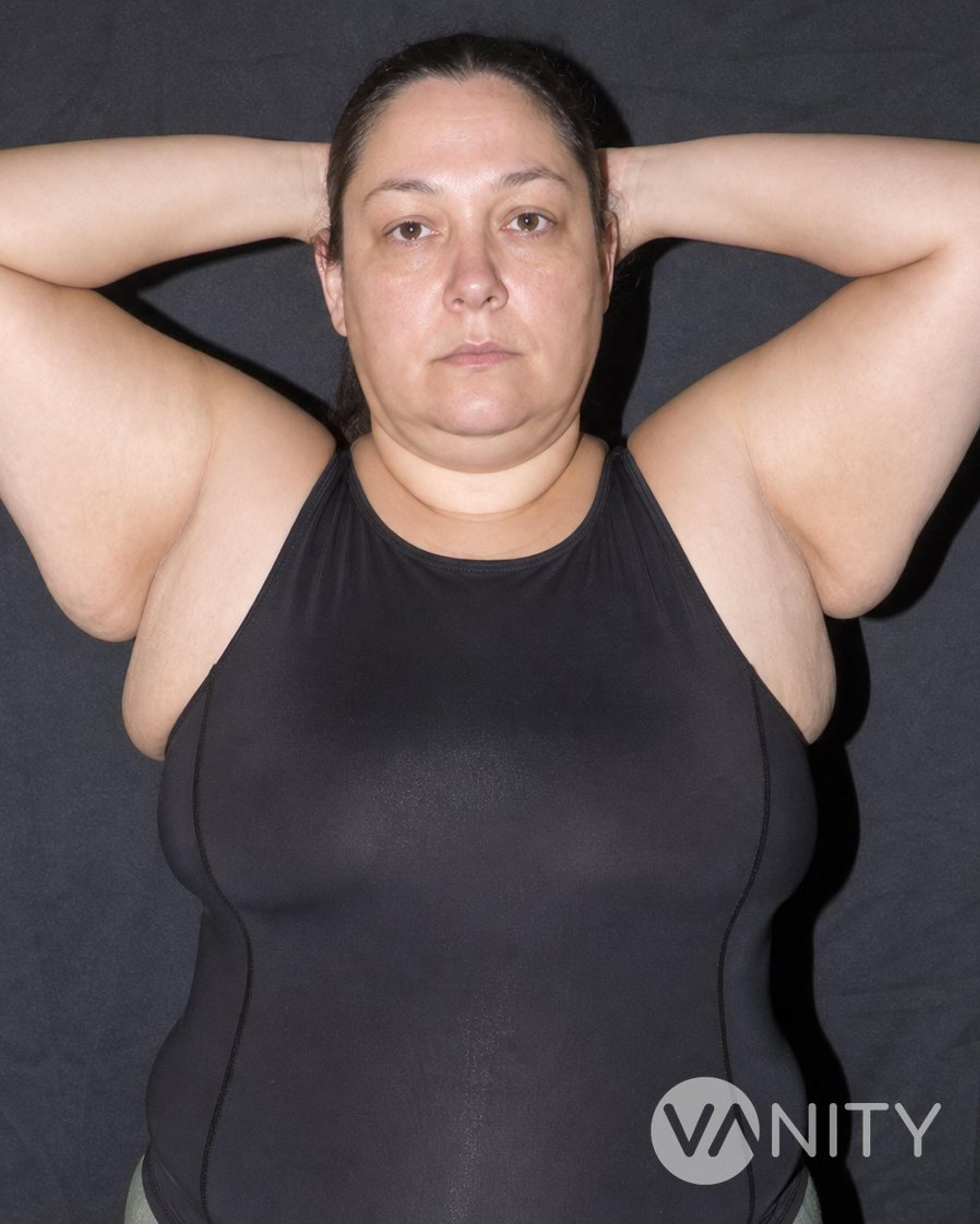
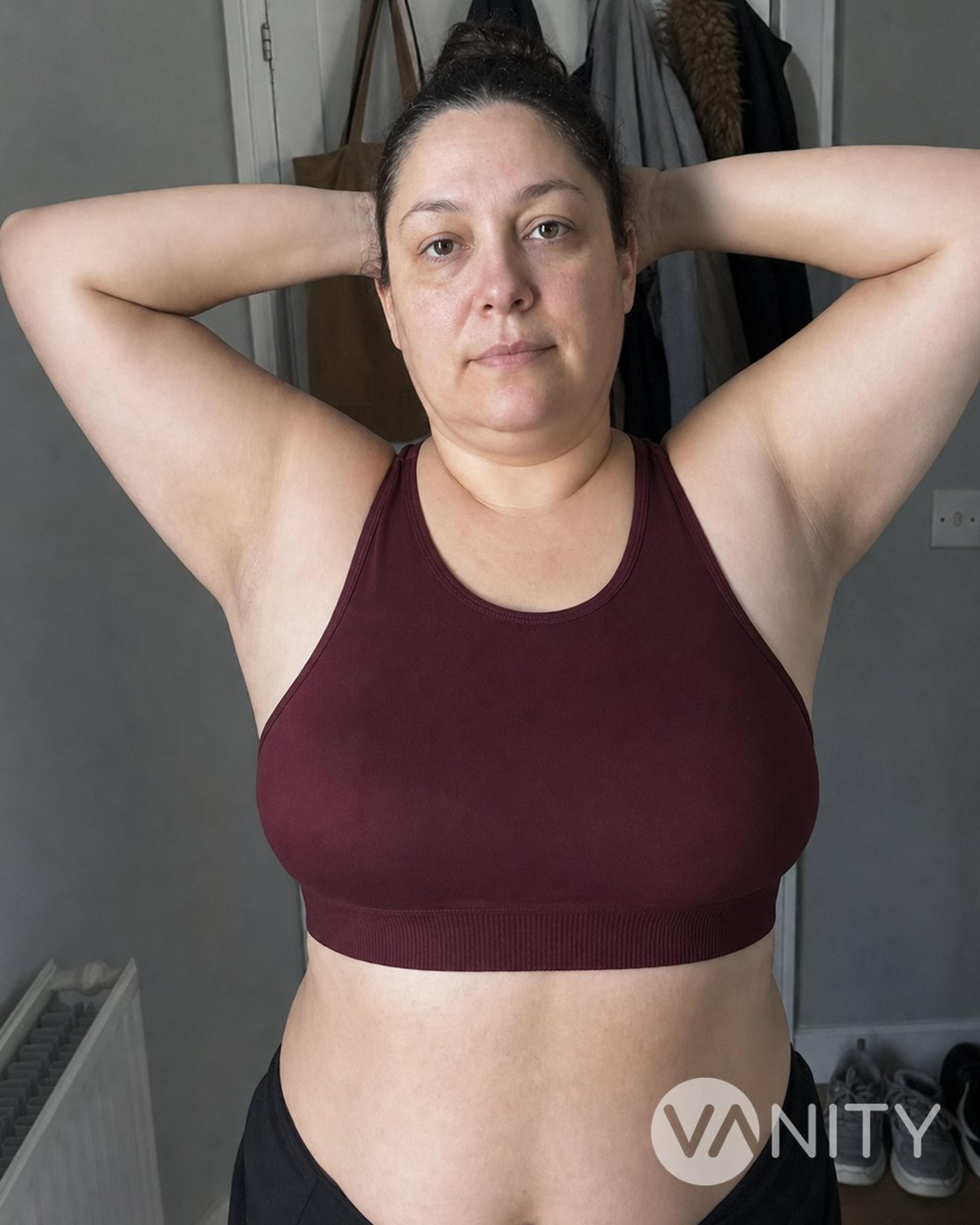
Accessory breast tissue is more common than many people realise and often appears on both sides. People usually come to us because of armpit pain that follows their cycle, a bulge that does not change with weight loss, discomfort when moving the arm, or because clothes do not sit comfortably. Any lasting lump in the armpit should be checked with imaging before treatment, as this is important for your safety.
Accessory breast removal: techniques we use
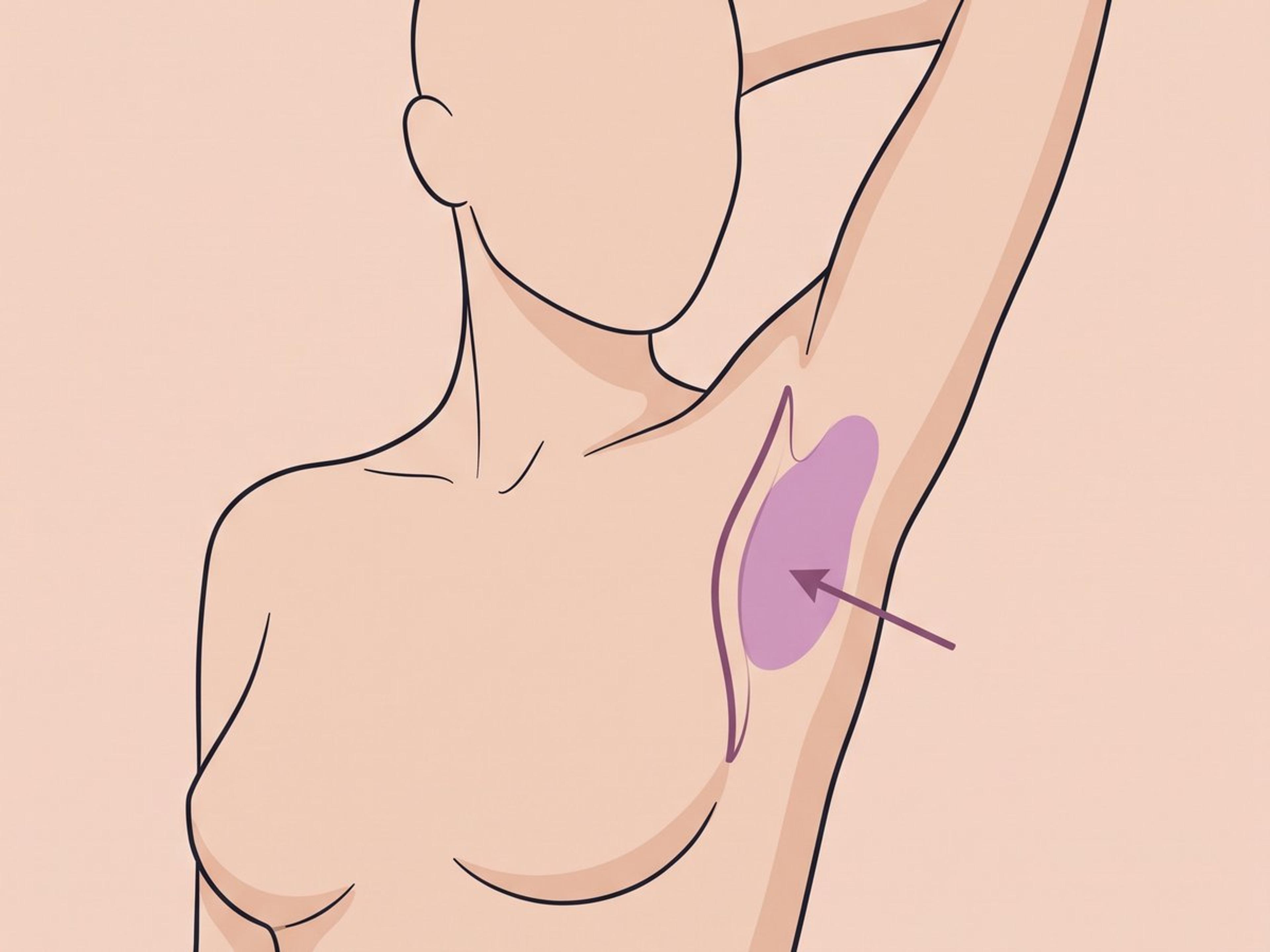
The breast tissue is removed through a small incision in the armpit and sent to the laboratory for testing. This is the most reliable way to remove breast tissue fully. It does leave a scar, and some fluid can collect under the skin while you heal.
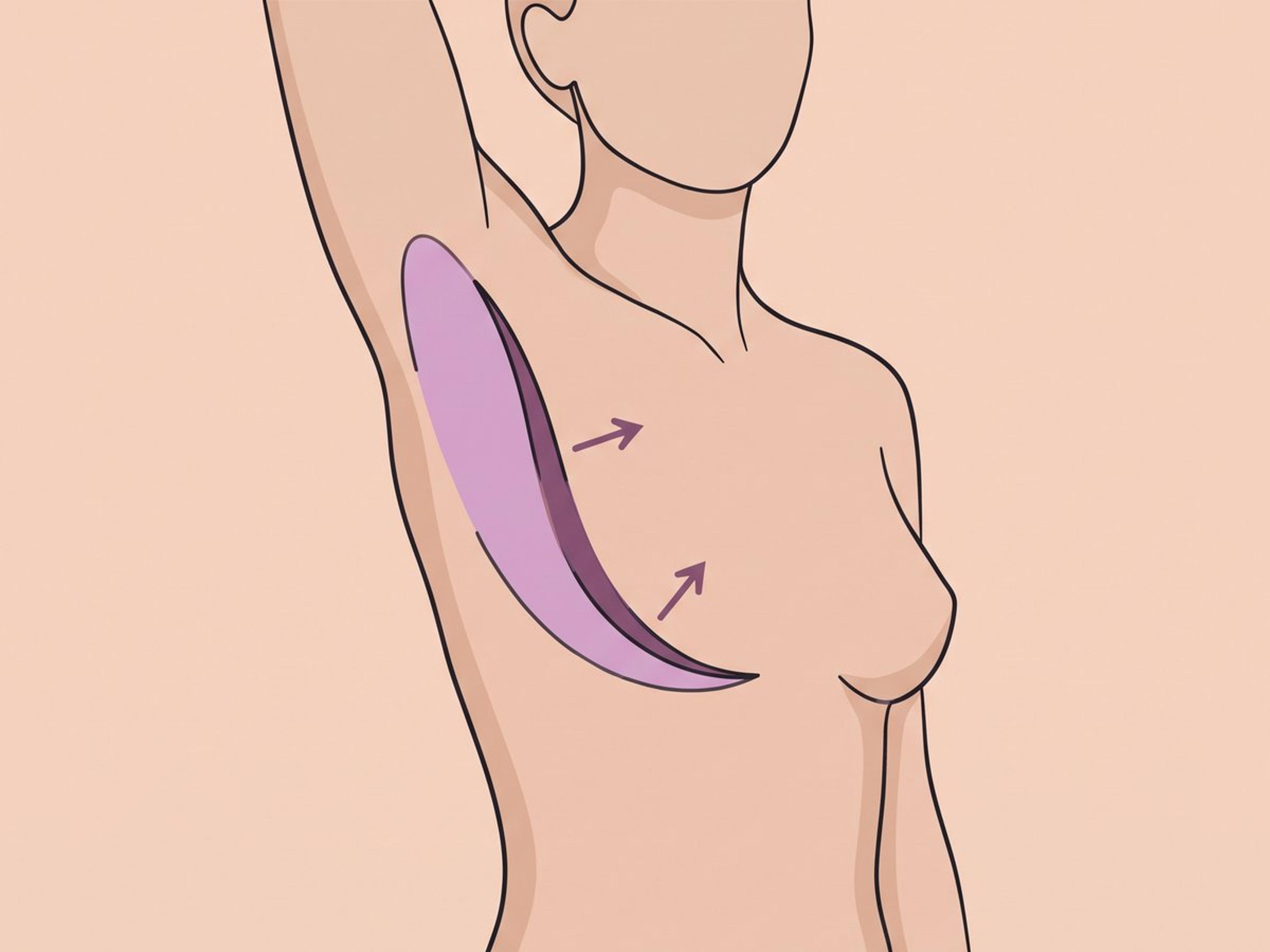
If there is loose skin as well as extra tissue, we remove a small section of skin, breast tissue and fat together. The incision follows the natural armpit fold to help the area sit smoothly when you move your arm. This option can improve loose skin in a way liposuction cannot.
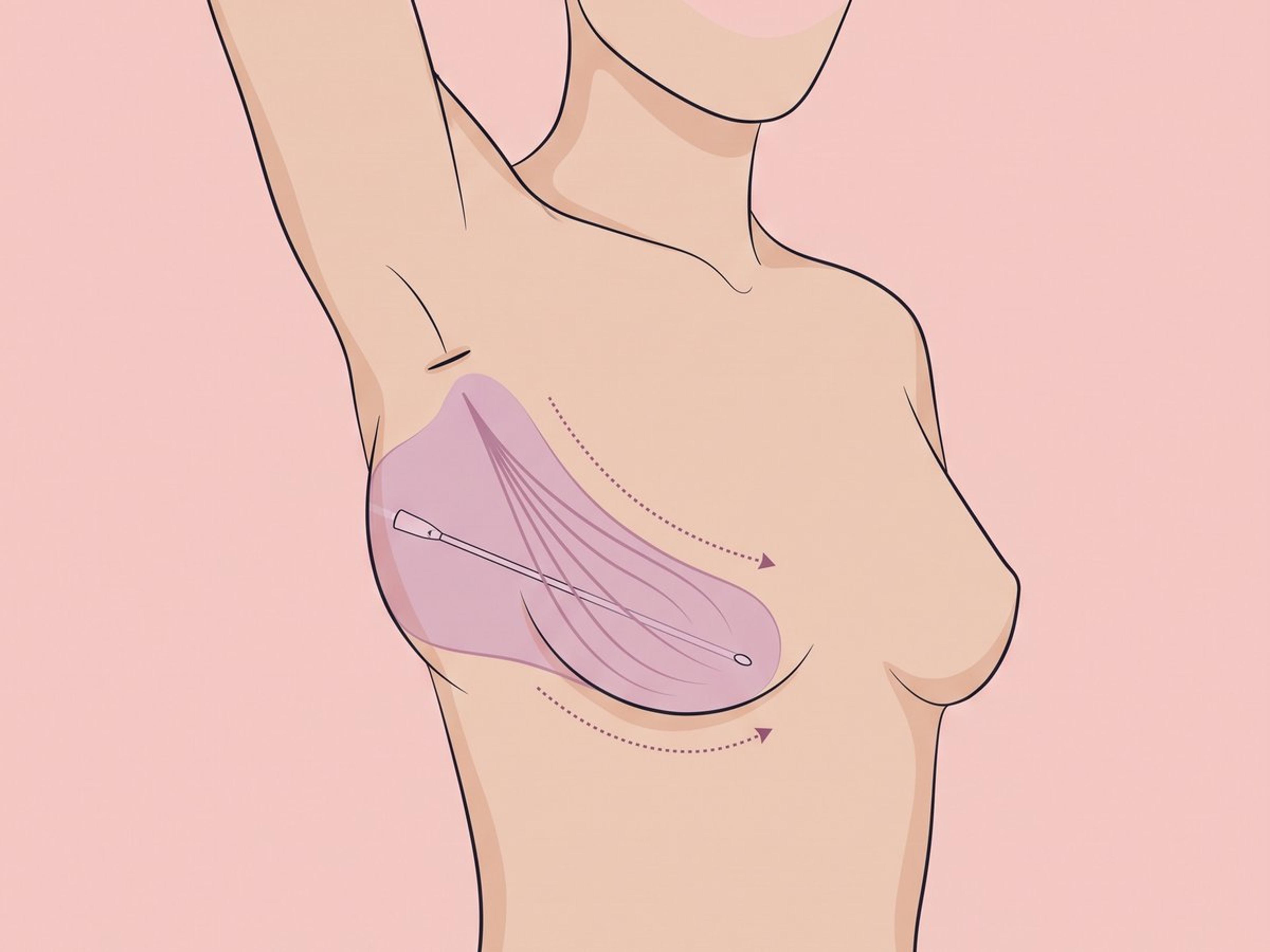
Liposuction removes extra fat through one or two very small openings, leaving minimal scarring. It is suitable only when the fullness is mainly fat. It cannot remove firm breast tissue or tighten loose skin, so some fullness or cycle-related tenderness may remain.
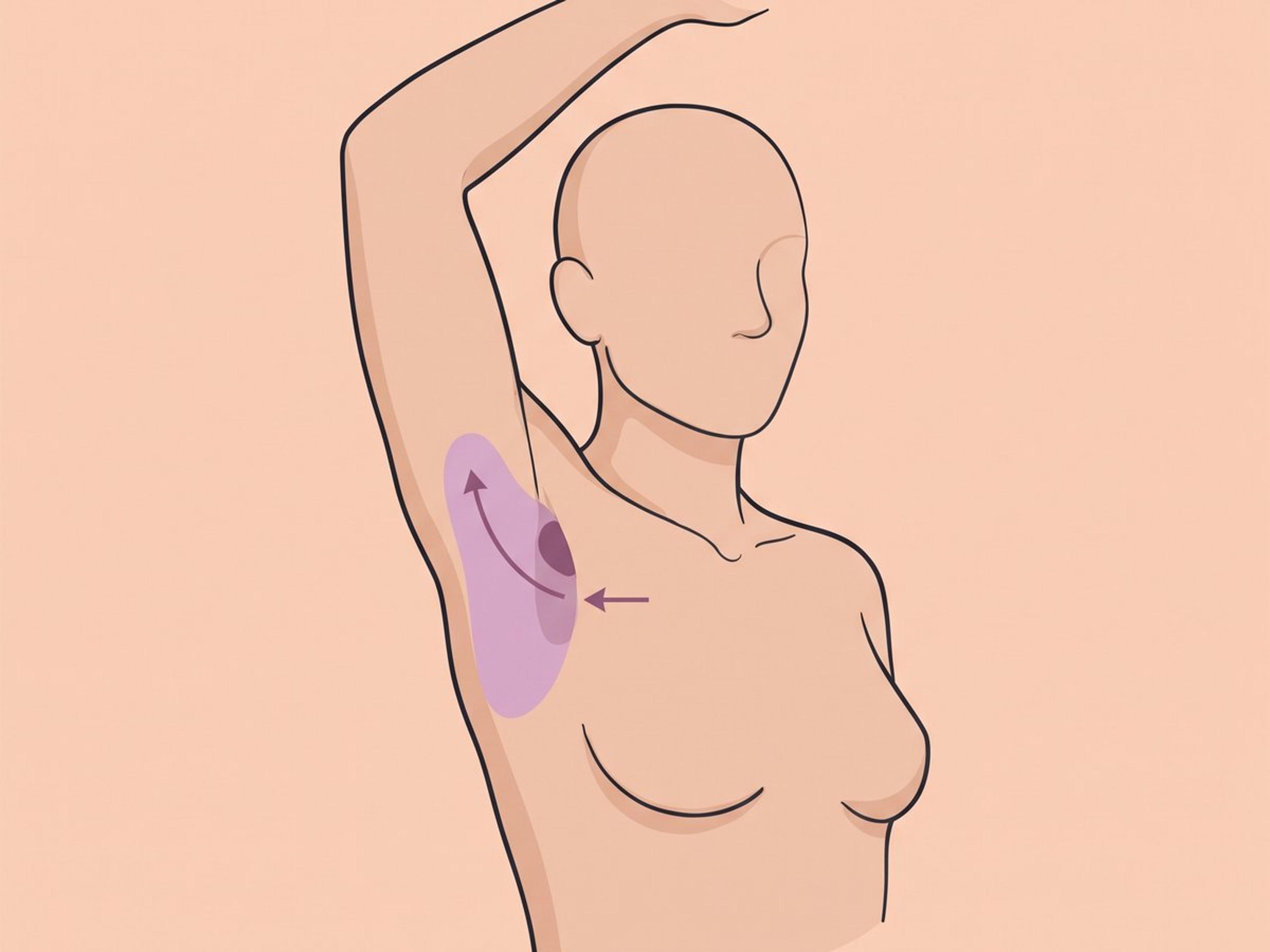
Liposuction removes the surrounding fat first. We then remove the remaining breast tissue through a short incision hidden in the armpit fold. This often offers a balance between a shorter scar and removing the tissue properly.
The operation, step by step
Standing marking, arm abducted
The borders of accessory tissue and the true degree of skin laxity are only definable upright with the arm raised — lying flat, the bulge flattens and the fold disappears. What is drawn here decides where the incision sits and whether skin has to come out.
Anaesthesia chosen by the size of the job
A small unilateral excision is done under local anaesthetic with sedation. Bilateral excision, and any case where skin is being resected, is more often done under general — because both axillae have to be worked on with the arms positioned, and that is not comfortable awake.
Liposuction takes the fatty envelope first
Tumescent or power-assisted liposuction through a stab incision removes the fat sitting over and around the gland. Debulking first shrinks what has to be delivered through the incision afterwards, which is what keeps the scar short.
The gland is excised through the axillary crease
The remaining fibroglandular mass is dissected free — usually down to the clavipectoral fascia — through a short incision hidden in the crease, staying clear of the axillary lymphatic channels and the intercostobrachial nerve. Liposuction cannot remove this component; leaving it behind is what produces residual bulk and continuing cyclical pain.
Dead space closed, specimen to pathology
Removing a mass leaves a cavity, and a cavity fills with fluid — so the space is obliterated with quilting sutures and a closed suction drain is placed where the dissection was wide. Seroma is the most frequent complication of this operation, and this step is the one that limits it. Every specimen goes for histopathology, because ectopic breast tissue carries the same malignant potential as the breast.
Compression on, home the same day
A compression garment goes on in theatre and stays on, holding the skin down onto the chest wall while the cavity seals. You go home the same day with drain-care instructions if one was placed, and gentle arm mobilisation starts early to reduce the chance of axillary cording.
Accessory breast removal: recovery week by week
- Day 1
The axilla is swollen, bruised and tight. A compression garment is worn, and a closed suction drain may be in place if a wide cavity was created. Raising the arm above shoulder height is restricted to protect the closure.
- Week 1
Any drain comes out and the first wound check happens. Seroma — the most frequent complication here, around 11% in large series and higher where drains are left out — usually declares itself in this window, and is managed with aspiration and continued compression.
- Weeks 2–3
Visible swelling largely settles and most people return to desk work and ordinary clothing. Altered sensation over the inner upper arm, from handling of the intercostobrachial nerve, may still be there. Axillary cording — a tight palpable band that limits how far the arm lifts — can appear now and usually resolves with physiotherapy.
- Week 6
Full shoulder range of motion and unrestricted exercise are usually allowed. The contour is broadly settled, and cyclical pain, where that was the presenting symptom, should have gone. Scars are at their reddest and firmest at this point.
- Month 12
Scar maturation is essentially complete and the contour is final. Scarring from accessory tissue removal can take up to 2 years to fade fully. Hypertrophic scars, reported in roughly 5–13% of cases, are treated with steroid injection or revision.
Accessory breast removal: how to prepare
- Have an axillary ultrasound — with mammography or MRI where indicated — if you feel a mass to confirm the mass is accessory breast tissue and not lymphadenopathy, a lipoma, or a malignancy, since ectopic glandular tissue can harbour carcinoma
- Defer surgery until several months after pregnancy and the end of breastfeeding, because accessory tissue enlarges and can lactate under hormonal stimulation
- Stop nicotine, and pause anticoagulants, antiplatelet agents and bleeding-risk supplements on surgical advice before the operation
- Attend a standing marking and photography session — the borders of accessory tissue and the degree of skin laxity are only definable upright, with the arm abducted
- Arrange a compression garment, drain care instructions if a drain is planned, and time away from overhead lifting and upper-body exercise
Accessory breast removal: risks
Every operation carries risk. We name them so you can ask the right questions.
- Seroma — the most common complication can be managed with aspiration and compression
- Axillary cording (axillary web syndrome) — reported in roughly 5–13% of cases, restricting shoulder abduction, usually resolving with physiotherapy
- Hypertrophic or widened scarring in the axilla — reported in about 5–13% of cases, treated with steroid injection or scar revision
- Temporary altered sensation over the inner upper arm
- Contour irregularity, dead-space depression, or incomplete removal — the characteristic failure of liposuction-only treatment of fibroglandular tissue, with cyclical pain persisting
- Hematoma, wound infection, and — where dissection disrupts axillary lymphatic channels — disturbance of lymphatic drainage

Accessory breast removal in Turkey: what to expect
This is day-case surgery, but the week after it is where the clinically useful events happen — drain removal, the first dressing change, and the point at which a seroma shows itself. We plan the trip around that week, not around the operating day.
Before you fly
Talk to the surgeon before you book anything. We want your imaging and your history first, because the technique is chosen from what the tissue actually is — predominantly fat, predominantly gland, gland with loose skin — and that decision changes the scar, the operating time, and whether a drain is used. Confirm in writing what the package covers and what it does not: histopathology of the excised tissue, aspiration of a seroma, and revision for hypertrophic scarring are the three worth naming explicitly.
At the clinic
Marking is done standing, with the arm up. Anaesthesia is general for most cases. Operating time for combined liposuction and minimal-incision excision averages over an hour. You can go home the same day in a compression garment, with a drain if a wide cavity was created.
Recovery in Istanbul
Plan for 7 days in Istanbul and 6 hotel nights, with 1 follow-up visits. Seroma is the most frequently reported complication of this operation — 6.25–37% across published series — and it tends to declare itself in exactly this window. Being here when it does means a needle aspiration in clinic rather than an emergency presentation at home. Compression stays on, and gentle arm mobilisation starts early, because axillary cording is a recognised early complication.
Heading home
Fly on day 7. The NHS and BAPRAS both advise against flying for 5 to 7 days after breast surgery and liposuction, and where a drain was used the flight follows drain removal and a wound check rather than preceding them. Stay in touch with us so we can follow up on your healing, our care continues for 12 months.
Checking a clinic in Turkey
Turkey rewrote its medical-tourism rules on 26 April 2025, replacing the 2017 regulation. Any facility treating international patients must now hold an international health tourism authorisation certificate, register on the Ministry of Health's HealthTürkiye portal, and carry complication insurance for the procedures it performs; hospitals and medical centres also had to complete TÜSKA accreditation by the end of that year. Facilities are required to publish that certification — so you can check ours, and you should check anyone else's before you pay a deposit.
Breast work is the area where a decision made in an afternoon is lived with for a decade, so press on the parts a brochure skips. Ask whether you are operated on in a hospital or a day unit and whether there is a bed for the night; ask for the named surgeon and their Turkish Medical Association registration; and ask what is written down if you need a revision after you have flown home. Where implants are involved, leave with their batch details in writing — they are what any surgeon anywhere will need later.
Most nationalities enter Turkey visa-free or on an e-Visa for a stay of this length, with a passport valid at least six months. Confirm the rule for your own passport, and build the return flight around the last clinic visit rather than fixing it in advance.
- United Kingdom
- £3,000 – £6,000
- United States
- $5,000 – $10,000
- Western Europe
- €3,000 – €6,500
Published typical ranges for orientation — confirm with a local quote.
Accessory breast removal: frequently asked questions
Is this just armpit fat, or is it actually breast tissue?
True accessory breast tissue is real breast parenchyma left over from the embryonic milk line, not simple fat. The clinical tell is hormonal behaviour: it swells and aches before a period, enlarges in pregnancy, and does not go away with weight loss. Ultrasound separates the two and decides whether liposuction alone can work.
Will liposuction alone remove it?
Only where assessment shows the excess is predominantly fat. Liposuction is documented as inadequate for fibroglandular tissue and does nothing for excess skin, so glandular cases treated this way tend to leave residual bulk and continued cyclical pain. Published treatment algorithms select the technique on tissue type, volume, skin laxity, and whether an accessory nipple is present.
Where is the scar and how visible is it?
Incisions sit inside the axillary crease or along a natural fold, so the scar lies in shadow with the arm down. Length depends on technique — a stab incision for liposuction, a crescentic resection where skin has to come out. Hypertrophic scarring is reported in roughly 5–13% of cases, and the axilla is a mobile, high-tension site, so scars can take a year or more to mature.
Does it come back after surgery?
Glandular tissue that has been completely excised does not regenerate. A bulge that returns usually means incomplete removal at the first operation — most often liposuction used on tissue that was glandular — or weight gain adding fat around the site. Histopathology of the excised specimen confirms what was actually taken out.
Can accessory breast tissue develop cancer?
Yes, though it is rare. Because ectopic tissue is true breast parenchyma, it can develop the same pathology as the breast, including invasive carcinoma, and case reports of this are well documented. That is why a persistent axillary mass warrants imaging before it is treated as a purely cosmetic problem, and why we send every excised specimen for pathology.
How long until I can use my arm normally?
Overhead reaching and lifting are restricted for the first 1 to 2 weeks, to protect the closure and reduce the risk of seroma. Full shoulder range of motion and unrestricted exercise are usually at around 6 weeks. If axillary cording develops it can limit abduction for a while; published series report it resolving with physiotherapy.
Is it done under general or local anaesthetic?
Both are used, and the choice follows volume, whether both sides are being treated, and whether skin is being resected. A small unilateral excision can be done under local anaesthetic with sedation, while bilateral excision with dermato-lipectomy is more often done under general. Reported operative time for combined liposuction and minimal-incision excision averages under an hour.
Who should not have this operation — or not yet?
Anyone pregnant, breastfeeding, or within a few months of stopping. The tissue is hormonally active and enlarges under stimulation, so we operate on a settled gland, not a swollen one. An axillary mass that has not been imaged is not operated on either — ultrasound comes first, to exclude lymphadenopathy and malignancy. Beyond that, the usual surgical rules apply: uncontrolled diabetes, a bleeding or clotting disorder, and anticoagulants that cannot safely be paused each defer surgery, as does active skin infection in the axilla — including a hidradenitis flare, which must be quiet before elective incisions are made there. Nicotine has to stop, and where liposuction is part of the plan, weight should be stable.
What should make me contact the clinic after surgery?
Spreading redness or warmth around the incision, discharge from the wound, fever above 38°C, or pain that gets worse after the first days instead of easing — those are the signs of infection, and they need review the same day, not watching. A sudden tense swelling in the armpit also warrants a call: in the first day or two that points to a hematoma; from the first week on it is more likely a seroma, the most common complication of this operation, dealt with by a needle aspiration in clinic. A tight cord-like band that limits how far the arm lifts is axillary cording — uncomfortable but not an emergency; raise it at follow-up and it usually resolves with physiotherapy.
Will I still need breast screening after the tissue is removed?
Yes. Excising an accessory focus removes that tissue — and we send every specimen to pathology — but this is not a preventive mastectomy, and it does not change your breast-cancer risk or your routine screening schedule for the breasts themselves. Nor can any operation guarantee that no microscopic ectopic tissue remains elsewhere along the milk line. Treat this as a contour and symptom operation with a tissue diagnosis built in, not as a cancer-risk procedure.
Will removing it affect breastfeeding later?
No. The operation is confined to the axilla and does not touch the ducts, nipple or parenchyma of the breast itself, so milk supply from the normal breast is unaffected. If anything it removes a future problem: accessory tissue engorges when the milk comes in and, having no outlet in most cases, can become painfully swollen for several days. The caveat runs the other way — we do not operate during pregnancy or breastfeeding, or for a few months after, because the tissue is hormonally swollen then and its true borders cannot be judged.
How much does accessory breast removal cost in Turkey?
€2,000 to €3,500 with us, confirmed after a consultation rather than quoted blind. For comparison it is typically £3,000 – £6,000 in the United Kingdom, $5,000 – $10,000 in the United States and €3,000 – €6,500 in Western Europe. What moves it is whether the tissue is taken by excision alone or with liposuction, and whether one axilla is treated or both.
Sources
- [1]Accessory Axillary Breast Excision with Liposuction Using Minimal Incision: A Preliminary Report · Aesthetic Plastic Surgery (PubMed/NLM) · 2017
- [2]Accessory Breast Tissue · Eplasty — PubMed Central · 2012
- [3]The ABCs of Accessory Breast Tissue: Basic Information Every Radiologist Should Know · American Journal of Roentgenology (PubMed/NLM) · 2014