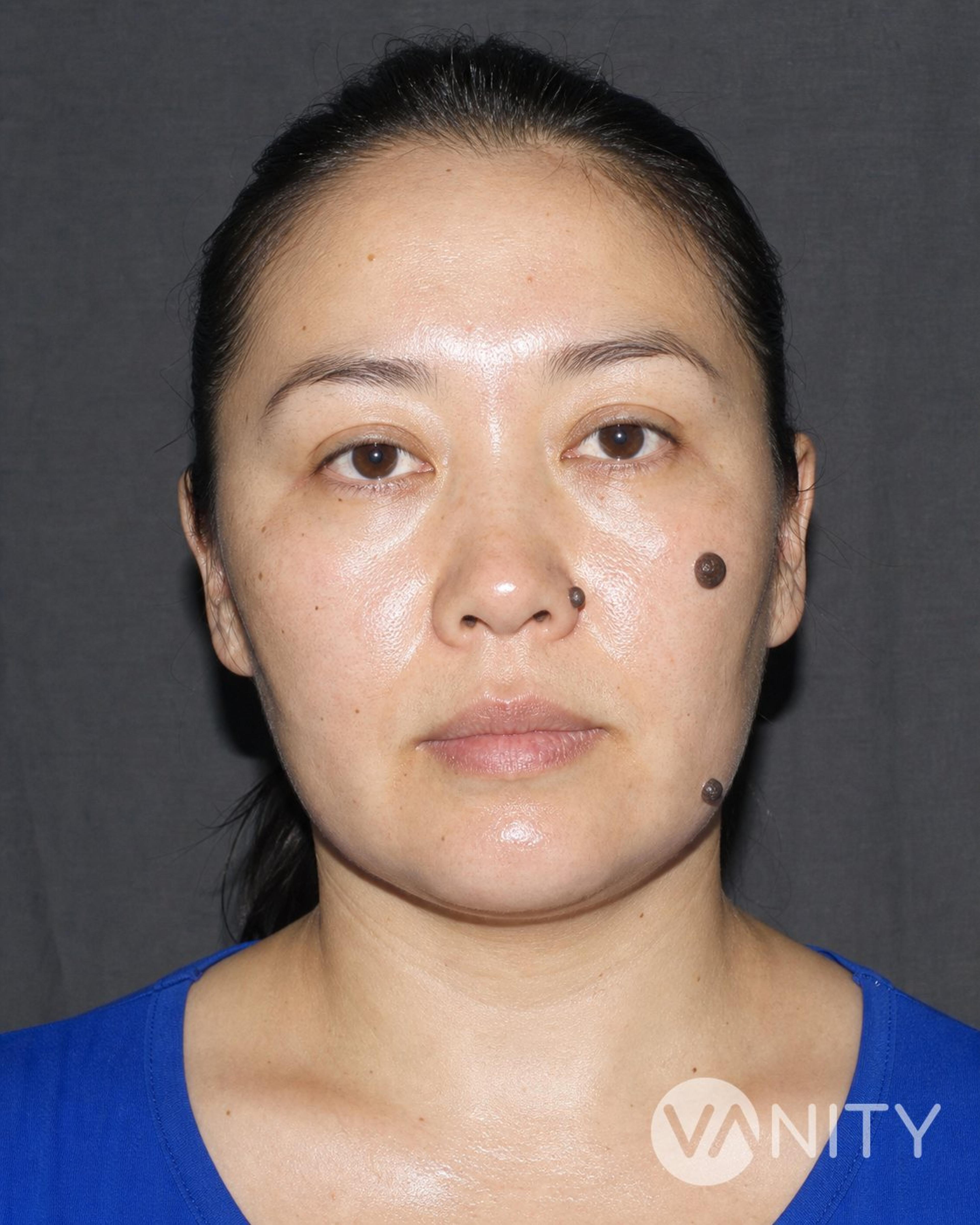
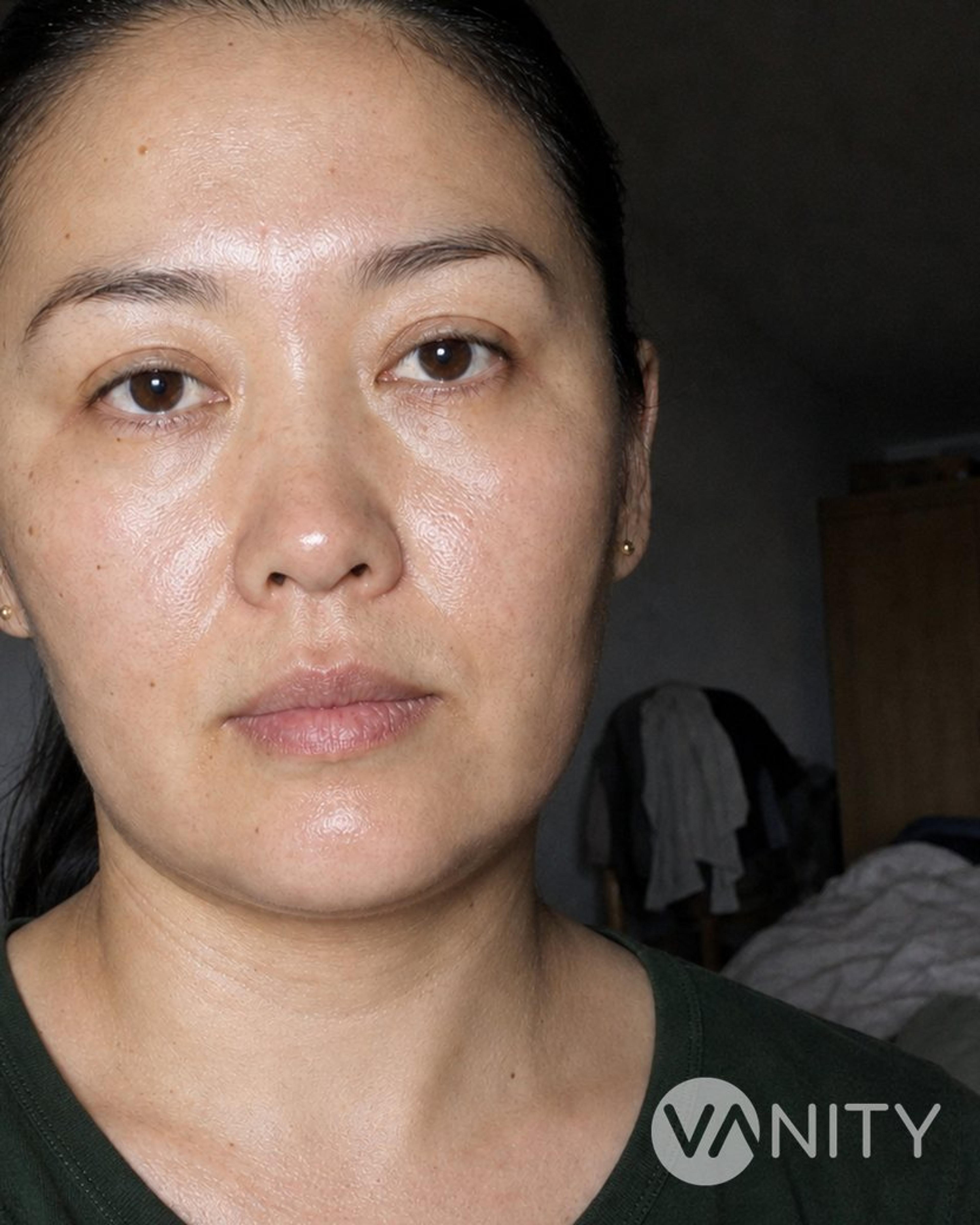



What it is
A mole is a cluster of pigment-making cells — melanocytes — sitting in the skin. Removing one is minor surgery done in a treatment room under local anaesthetic: the lesion is either shaved off flush with the surrounding skin and the base sealed, or cut out full-thickness with a scalpel and closed with sutures. Either way the tissue goes to a pathologist, who examines it under a microscope for atypical or cancerous cells. That examination is as much the point of the procedure as the removal is, which is why we do not burn, freeze or laser a pigmented mole — those methods leave nothing to look at.
Three situations bring people to us. A mole that has changed in size, shape or colour and needs a diagnosis. A mole that catches on a bra strap, a waistband or a razor. And a mole you simply do not want, which is a reason on its own — cosmetic removal is a recognised indication, usually a private one. If you have many moles, or atypical ones, the right answer is generally photography and surveillance rather than removing them all: taking out one mole does not lower your melanoma risk anywhere else on your skin.
Techniques we use
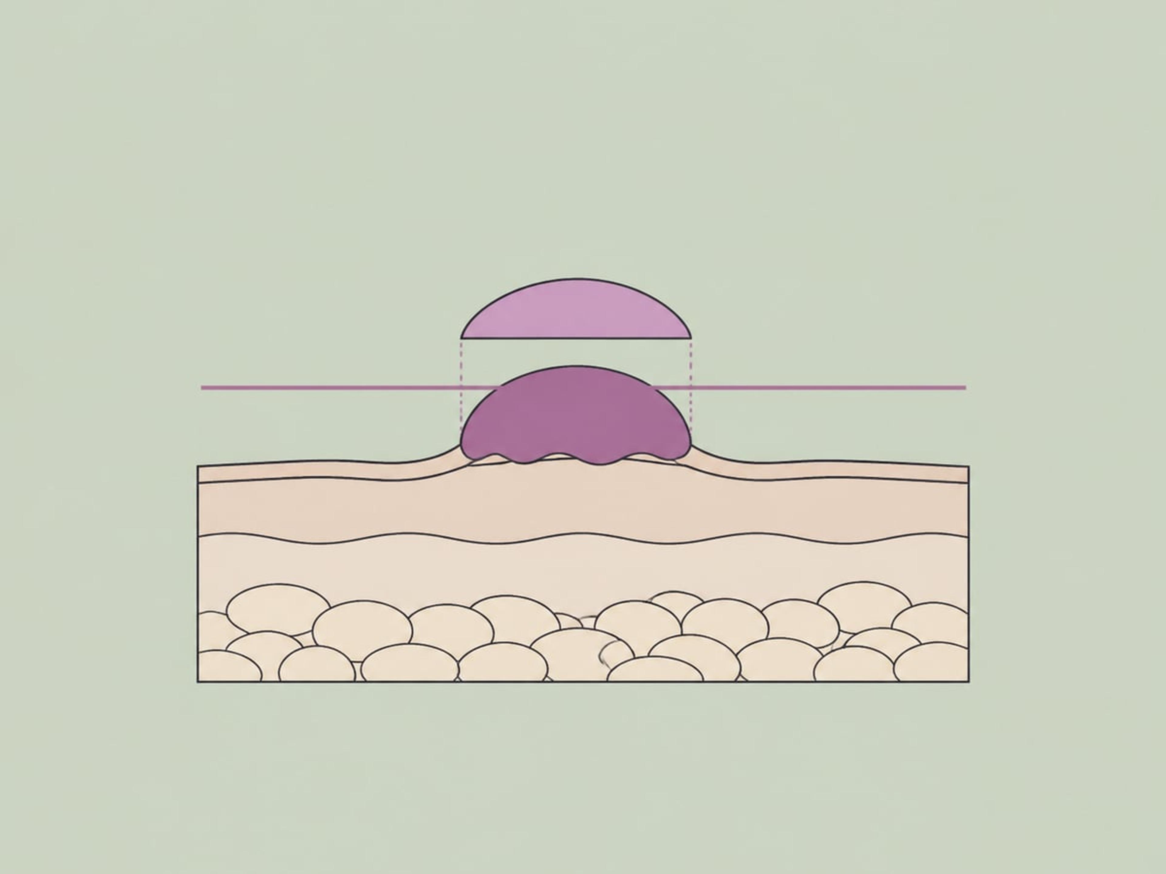
A blade passes tangentially through the mole and takes it off level with the surrounding skin; the base is then sealed with electrocautery or a chemical haemostatic. There are no sutures and the wound heals open, which gives a good contour on raised moles and still leaves a specimen for the laboratory. Because the deeper dermal part of the naevus stays behind, pigment returns in the scar more often than after full excision.
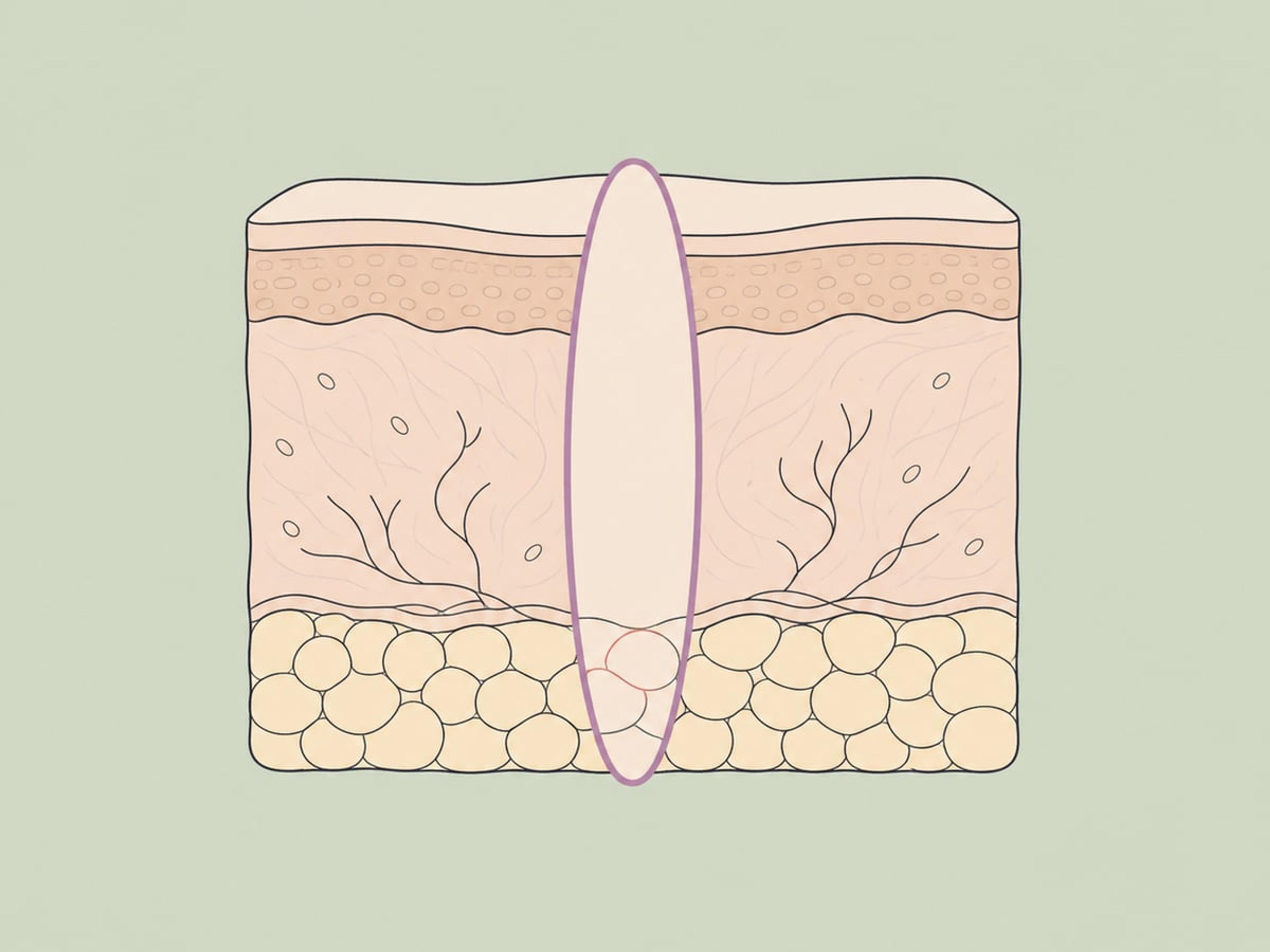
The mole is cut out through the whole thickness of the skin, usually as a narrow ellipse with a small margin of normal skin around it, and closed with sutures. The entire lesion including its dermal component goes to histopathology, which makes this the method we choose whenever anything about the mole looks suspicious. The trade is a linear scar noticeably longer than the mole it replaced.

A small circular cutting blade is pressed through the skin around the lesion and lifts out a full-thickness cylinder of tissue, closed with one or two sutures. It suits small, well-defined moles and lesions with a deep dermal component. It sits alongside excisional biopsy as a standard way of sampling a mole that needs a diagnosis.
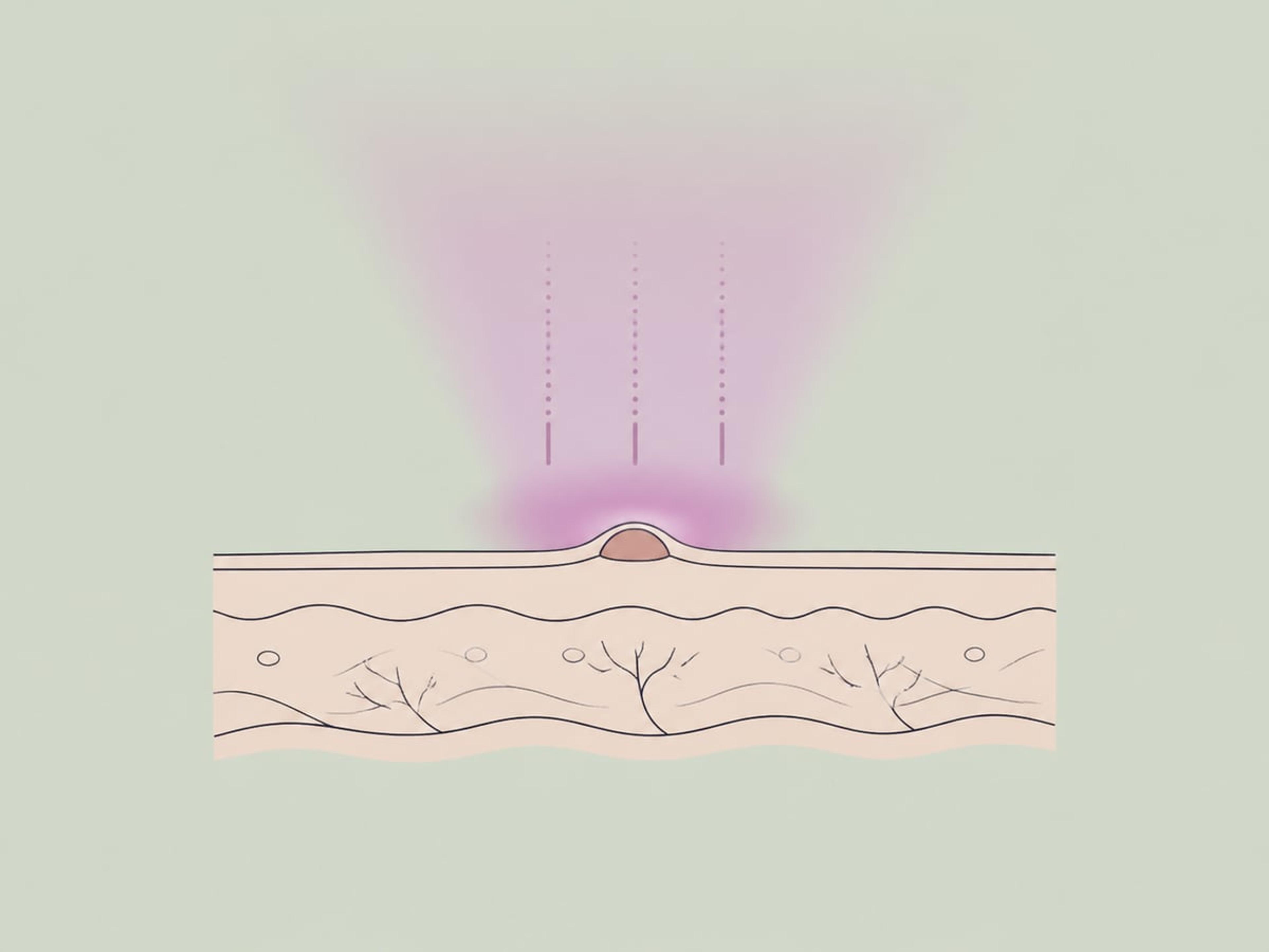
These destroy the lesion with heat or cold rather than removing it intact, so nothing survives to send to a pathologist and recurrence rates are higher. We do not use them on pigmented moles, and the British Association of Dermatologists advises surgical excision for that exact reason. Melanomas have been reported in laser-treated sites 1 to 10 years afterwards, with no pre-treatment histology on record.
The removal, step by step
Dermoscopy first, then the plan
The mole is examined under dermoscopy before anything is decided, because how deep the pigment sits and how ordered its pattern looks are what choose the method — a raised, plainly benign mole suits a shave, while anything with an irregular network is taken full-thickness so the pathologist sees the whole lesion. The plan is settled in front of you, at the skin, not booked in advance from a photograph.
Numbing the site
The area is numbed with a topical agent and then injected local anaesthetic, usually with adrenaline, which narrows the small vessels so the field stays dry and the block lasts through the removal. The injection stings for a few seconds; after that the removal itself should be felt as pressure and nothing more. You stay awake, and you can drive yourself home.
The mole comes off
A shave excision passes a blade tangentially through the lesion and takes it off flush with the surrounding skin, which gives the better contour on raised moles. A punch or elliptical excision instead removes the full thickness of skin with a narrow margin of normal tissue around it — the choice whenever the lesion needs a definitive answer rather than a tidy result. One lesion takes 15 to 30 minutes.
Sealing or closing the wound
A shave base is sealed with electrocautery or a chemical haemostatic and left to heal open, which is why it leaves a flat pale circle rather than a line. An excision is closed with sutures placed along the skin's relaxed tension lines, because a scar that runs with the natural creases is pulled on least and stays narrowest.
The specimen goes to the laboratory
The tissue is labelled at the treatment couch, fixed, and sent to a pathologist who examines it for atypical or cancerous cells. This is the step that outlasts your trip: reporting takes several working days, so ask in which language the report is written and for the block and slide accession numbers, which let your own dermatologist request a second review anywhere in the world.
Recovery, week by week
- Day 1
The dressing comes off the day after. From then on the wound is washed gently with mild fragrance-free soap, patted dry, covered with petroleum jelly from a squeeze tube and re-dressed daily — we skip antibiotic ointment, because contact allergy to it is a likelier problem than infection. Stinging or burning at the site is expected; most people are back at work the same day.
- Week 1
Shave and punch wounds close over at 7 to 10 days, and facial sutures come out at 5 to 7. This is the window in which anything wrong declares itself: pain should be fading, not building, and spreading redness, thick yellow discharge or fever means messaging us with photographs rather than waiting it out. Showering is fine with a fresh dressing after; nothing submerged.
- Weeks 2–3
Sutures on the trunk and limbs come out at 10 to 14 days, usually at home, so arrange that before you travel. Once the wound is closed — commonly 1 to 2 weeks — pools and hot tubs are back on. Strenuous exercise waits until the sutures are out if the site was a mobile, high-tension one such as the shoulder, back or knee, since pulling on a fresh line is what widens a scar.
- Months 1–3
The site is healed and covered in new skin that looks pink or red — this is the scar at its most obvious, not its final state. Broad-spectrum SPF 30 or higher over it every day genuinely changes the outcome, because fresh scar tissue darkens with sun and post-inflammatory pigmentation is slow to fade, more so in darker phototypes.
- 6 months
If pigment is going to return in a shave-excision scar it typically shows around now, averaging about five and a half months — the recurrent naevus phenomenon, and a reason to have any new colour looked at rather than assumed benign. Pigment spreading beyond the scar border is the finding that matters most.
- 12 months
Redness and firmness have settled and the scar has reached its final appearance. What you can see at a year is what you keep.
How to prepare
- Have the mole examined with dermoscopy before you agree to remove it, and bring any older photographs of it so change over time can be judged rather than guessed.
- Tell us about every anticoagulant, antiplatelet drug, NSAID and supplement you take, and any bleeding disorder — and do not stop a prescribed medicine without asking the doctor who prescribed it.
- Tell us if you have ever formed keloid or hypertrophic scars, reacted badly to a local anaesthetic, or had a cardiac device implanted, since electrocautery is part of the procedure.
- Confirm before the day that the lesion will be sent for histopathological examination, and decline laser, cautery or cryotherapy for a pigmented mole — those leave no tissue to examine.
- Do not use over-the-counter mole or skin-tag removal creams, pens or patches. No such drug product is approved, and the reported harms include skin injury, infection needing antibiotics, severe scarring and a skin cancer diagnosed late because it was mistaken for a harmless mole.
Risks
Every operation carries risk. We name them so you can ask the right questions.
- A scar at every removal site, including hypertrophic or keloid scarring, which is more common on the chest, shoulders and upper back. Scarring is certain rather than possible — the only question is how visible it is.
- Bleeding during the procedure or in the first 24 hours, made more likely by anticoagulant and antiplatelet medication.
- Wound infection, running at roughly 1 to 2% in dermatologic surgery without prophylactic antibiotics, higher below the knee and lower on the face. It shows as increasing pain, spreading redness or warmth, pus, yellow crusting or fever.
- Incomplete removal, with residual dermal melanocytes repigmenting the scar — the recurrent naevus phenomenon. Histologically confirmed recurrence or persistence after shave removal of mildly atypical dysplastic naevi is around 10% at 2 years, averaging 5.5 months to appear, and is strongly associated with the lesion extending to the lateral or follicular margins of the original specimen. The regrowth can mimic melanoma closely enough to need re-excision to tell them apart.
- A delayed or missed skin cancer diagnosis when a lesion is destroyed without histopathology, or removed with an unapproved at-home product.
- Post-inflammatory hyperpigmentation or hypopigmentation at the treated site, more pronounced in darker skin phototypes, and, rarely, injury to a superficial cutaneous nerve leaving altered sensation.

What to expect in Turkey
Mole removal is a day case. You are not admitted, you are not sedated, and the visit turns on two short appointments — one to examine and remove, one to check the wound. The part that outlasts your trip is the pathology report.
Before you fly
Two things are checkable from home, and worth checking. In Turkey only physicians licensed under Law No. 1219 may perform this — in practice a dermatologist or a plastic, reconstructive and aesthetic surgeon; nurses and non-physician aestheticians hold no independent authority for it. Separately, the facility must hold an International Health Tourism Authorization Certificate under the 2017 Regulation on International Health Tourism and Tourist Health, and any agency arranging your care must be authorised by USHAŞ. Send us photographs and talk to the doctor before you book a flight.
At the clinic
The visit starts with an examination under dermoscopy, and the method is decided there, in front of you: shave, punch or full-thickness excision, depending on how the lesion sits in the skin and how suspicious it looks. The site is numbed, the removal takes 15 to 30 minutes for one lesion, and the specimen is labelled and sent to pathology. You are not kept in. You walk out, and you can drive yourself and go back to work the same day.
Recovery in Istanbul
Plan on 3 days in Istanbul and 2 hotel nights, with 2 appointments inside that window. The dressing comes off the day after the procedure; from then on the wound is washed gently with mild fragrance-free soap and water, patted dry, covered with petroleum jelly from a squeeze tube and re-dressed daily. We do not routinely use antibiotic ointment — contact allergy to it is a bigger risk than infection is. Expect stinging or burning at the site for a few days. Shower if you like, with a fresh dressing afterwards; no pools, no hot tubs, nothing submerged.
Heading home
There is no flying restriction after mole removal. The usual advice to wait about 10 days applies to chest and abdominal surgery, where cabin pressure and clot risk are real concerns — this procedure enters no body cavity, uses no general anaesthetic, leaves no drain and immobilises nobody. Flying the same day is safe; we prefer 2 days only so the wound can be inspected once and any bleeding handled here rather than in a departure lounge. Sutures come out at around 5 to 7 days on the face and 10 to 14 on the trunk and limbs — most international patients have that done at home, so arrange it before you travel and take the suture details with you. Pathology takes several working days, which means the report will usually reach you after you have landed: ask in which language it is written, how it will be sent, and for the block and slide accession numbers, so your own dermatologist can request a review.
- United Kingdom
- £200 – £600
- United States
- $150 – $1,500
- Western Europe
- €200 – €600
Published typical ranges for orientation — confirm with a local quote.
Frequently asked
Will a mole grow back after it is removed?
It can. Shave excision leaves the deeper dermal part of the naevus behind, so pigment sometimes reappears inside the scar, typically around 6 months later — the recurrent naevus phenomenon. Full-thickness excision recurs less often. Any regrowth should be looked at promptly, because pigment extending beyond the scar border is the strongest clue that the lesion is a melanoma rather than a benign recurrence.
Does mole removal leave a scar?
Yes. Anything that wounds the skin leaves a mark, and the trade differs by method: shave excision usually leaves a small flat pale or pink circle, while excision leaves a linear scar longer than the original mole. We plan the orientation and the closure to keep it quiet, but no technique is scar-free and we will not tell you otherwise.
Is the mole sent to a laboratory?
Yes. After both shave and surgical excision the tissue is examined under a microscope by a pathologist to check for atypical or cancerous cells, and the result is reported back to you. This is the main reason we avoid laser, cautery and cryotherapy for pigmented moles — those methods leave nothing to examine.
Does mole removal hurt?
The site is numbed with topical and injected local anaesthetic, so the removal itself should not be painful, though the injection stings briefly. Stinging or burning at the site for a few days afterwards is expected and simple painkillers usually cover it.
How long does the site take to heal, and when does the scar settle?
Most shave and biopsy wounds close within 7 to 10 days, and the site is generally healed at 2 to 3 weeks, leaving pink or red new skin. Redness fades over the following months, and broad-spectrum SPF 30 or higher over the healed site reduces the discolouration that follows — which is more pronounced in darker skin phototypes. The scar reaches its final appearance at about 12 months.
Can I have several moles removed in one visit?
Yes. Several lesions can be removed in the same sitting, each under its own local anaesthetic, and the per-lesion price typically falls when they are done together. Mole removal is a single session, not a course — if a provider quotes you a multi-session package, they are almost certainly describing laser or ablative destruction, which leaves no specimen for histology.
Are at-home mole removal creams and pens safe?
No. There are no approved prescription or over-the-counter drug products for removing moles, and what is sold for the purpose often contains high concentrations of salicylic acid or similar caustic agents. Reported harms include skin injury, infection requiring antibiotics, severe scarring, and delayed diagnosis of a skin cancer that was mistaken for a harmless mole.
When can I swim or exercise again?
Showering is fine, with a fresh dressing applied afterwards, but swimming pools, hot tubs and submerging the wound are out until it has healed — usually 1 to 2 weeks. After an excision on a mobile or high-tension site, hold off on strenuous activity until the sutures come out, so the wound does not pull open or the scar widen.
Is there anyone who shouldn't have a mole removed?
Almost anyone can, because the procedure needs only local anaesthetic — but a few situations change the plan. An active skin infection at or near the site means we postpone until it has cleared. Anticoagulants, antiplatelet drugs and bleeding disorders are managed rather than a bar: tell us about them, and never stop a prescribed medicine without the prescriber's agreement. A history of keloid or hypertrophic scarring is a reason to think hard before removing anything purely cosmetic from the chest, shoulders or upper back, where those scars form most readily. In pregnancy, moles commonly darken and enlarge slightly as a normal hormonal change, so we defer cosmetic removal until afterwards — though a genuinely suspicious lesion is still examined and, if necessary, biopsied promptly, pregnant or not, since the local anaesthetic doses used here are considered safe. And a true allergy to local anaesthetic, rare as it is, is something we need to know before the day, not during it.
What should make me contact the clinic after a mole removal?
Five things, and none of them should be waited out. Pain that increases after the first two or three days instead of fading. Redness or warmth spreading outward from the wound edge. Pus or thick yellow discharge — different from the thin, clear ooze a healing wound normally produces. Fever. And bleeding that does not stop after 15 to 20 minutes of firm, uninterrupted pressure. Most of our international patients are home before any of these could appear, which is exactly why we ask you to send photographs by message rather than wonder: a wound infection caught early is a short course of antibiotics; one caught late is a worse scar.
Can cutting a mole make it spread or turn cancerous?
No — this is one of the most persistent myths in dermatology, and it has the danger backwards. Cutting into a benign mole does not transform it into cancer, and excising a melanoma does not spread it; complete excision is precisely how melanoma is treated. The genuine risks sit elsewhere: destroying a mole with laser, cautery or cryotherapy leaves nothing for the pathologist to examine, and pigment returning in a shave-excision scar can mimic melanoma closely enough to need re-excision to tell them apart. The thing to fear is not the scalpel — it is the mole that was never examined at all.
Sources
- [1]5 reasons to see a dermatologist for mole, skin tag removal · American Academy of Dermatology (aad.org) · 2022
- [2]Moles · NHS (nhs.uk) · 2025
- [3]Prospective study of wound infections in dermatologic surgery in the absence of prophylactic antibiotics · PubMed, US National Library of Medicine · 2006
- [4]Mole Removal: Surgery, Aftercare & Scars · Cleveland Clinic (my.clevelandclinic.org) · 2025